- Abstract
Streptomyces species are most known for their antibiotics production. In this paper Streptomyces production of antibiotics was investigated in two sections. Firstly, Streptomyces was isolated from soil sample; their production of antibiotics was examined by agar plug diffusion and it has shown very poor antibiotic activity against three different indicator bacteria strains M. luteus, E. coli DS941, and E. coli DS941 PSB1C3. It is suggested that standardization of sample collection could optimize the probability of finding streptomyces strains that produce significant antibiotics. The soil sample was identified by 16S rRNA sequencing and found to be very closely related to Streptomyces brevispora strain BK160. Secondly, chloramphenicol production was investigated in two strains of Streptomyces venezuelae. It is shown that the genetically modified S. venezuelae M1582 strain has more efficient antibiotic production compared to the wild-type S. venezuelae strain ATCC10712, and that is due to its enhanced chloramphenicol gene cluster expression by improved sven0913 gene expression.
- Introduction
Antibiotics are compounds produced by bacteria against other types of bacteria as a defense or offense action (Hutchings et al., 2019). These antimicrobial compounds have been discovered and started to be used as drugs to cure many types of diseases in late 19 century. Since then, medicine has successfully conquered many infectious diseases, and thus have significantly reduced death rate, which was previously high due to the spread of infectious diseases such as tuberculosis, diarrhea, pneumonia, …etc. (Zaffiri et al., 2012). However, throughout the past years, there has been an over production and usage of antibiotics, leading bacteria to develop resistance against antibiotics. Moreover, some strains of bacteria have acquired multiple drug resistance (MDR) against more than one type of antibiotic, hence, became harder to kill. Antibiotic resistant bacteria are a great threat to the global health systems (Huemer et al., 2020).
Along with the growing concern about antibiotic resistant bacteria, many efforts are being put to reduce their risk by finding alternatives for antibiotics. Researchers have been exploring soil bacteria hoping that it might hold potential novel antibiotics to be discovered (D’Costa et al., 2007).
Streptomyces species isolated from soils are being intensively studied. Streptomyces is a genus of filamentous, spore forming, gram positive bacteria. Given their high abundance and prevalence in soils, in addition to their useful metabolites, Streptomyces have attracted so much attention. They are considered the main source of antibiotics as they’ve been used to produce 80% of all antibiotics (de Lima Procópio et al., 2012). Streptomyces produce variety of antibiotics. Their antibacterial mode of action against other species is by interruption protein synthesis by blocking their 30S ribosomal subunits. (Nicolaou and Rigol 2017)
This paper seeks to investigate the production of antibiotics from soil isolates. The experiment is divided into two sections, the first is to isolate an antibiotics-producing Streptomyces from soil, test its antibiotic production against different indicator bacteria strains and to identify it by 16S rRNA sequencing.
The second section is to examine the production of chloramphenicol antibiotics from wild-type S. venezuelae ATCC10712 and genetically modified M1582 S. venezuelae. In research by Fernández-Martínez and team, it was shown that in S. venezuelae a gene cluster for 9 proteins encoded by genes sven0916–sven0929 is required for Chloramphenicol production, three more genes were suggested to have a role in the production of Chloramphenicol including gene sven0913 which acts as a transcriptional activator for Chloramphenicol gene cluster. The researchers modified wild-type S. venezuelae to create M1582 strain which have a highly expressed sven0913 gene to enhance the activity of Chloramphenicol gene cluster (Fernández-Martínez et al., 2014)
- Methods
- Streptomyces from soil samples
For the first section of this study, Streptomyces was isolated from soil, to test their antibiotic production and identify it by 16S rRNA sequencing as described in lab manual (Colloms 2021)
- Soil sample and colony count
Soil sample was collected from Glasgow University campus (kelvin building). The sample was suspended in ringer’s solution, heat treated to kill non-spore forming species. And diluted in a serial dilution.
Samples were plated into two plates: Cycloheximide, and Cycloheximide + Nalidixic acid plate, and incubated. Then, number of colonies was counted to find colony-forming unit (CFU) per gram.
- Streaking soil samples into single colonies
To obtain pure isolates, two different suspected Streptomyces colonies based on their morphological characteristics (white, hard, powdery dry looking) were streaked in a Soya Flour mannitol (SFM) and incubated.
- Spreading on spore-forming plates
To enhance sporulation, two sporulation plates were used: Soya Flour mannitol (SFM), and Maltose yeast extract malt extract (MYM). Sample were plated in SFM and MYM plates and incubated for a week.
- Agar plug diffusion to check antibiotic formation
To test antibiotics formation, agar plug diffusion technique was used against three indicator bacteria: M. luteus, E. coli DS941, E. coli DS941 PSB1C3. Agar plugs from SFM and MYM soil samples were placed in a plates of each indicator bacteria, and plates were incubated.
- 16S rRNA amplification and visualizing
- PCR
To amplify 16S rRNA, a small colony from each soil sample was resuspended and heat treated to disrupt the cells and release their genomic DNA to be used as a templet for PCR. Samples were transferred to a walled PCR tube and added to it a mixture of master mix, primers, sterile distilled water, and DMSO. A no-templet control was added. And PCR was run.
The sequence of primers used for PCR:
27F 5’- AGA GTT TGA TCM TGG CTC AG –3’
519R 5’- GWA TTA CCG CGG CKG CTG -3’
- Gel electrophoresis
To assess 16S rRNA amplification, samples were run in 1% agarose gel electrophoresis, , and they were loaded with 1kb ladder. Bands were visualized under UV-light.
- 16S rRNA sequencing
Sample 2 was chosen for sequencing. Qiagen PCR clean up kit was used to prepare the sample and remove unwanted PCR left out components. Nanodrop UV spectrophotometer was used to measure the concentration of soil sample, and the sample was sent for sanger sequencing.
- Chloramphenicol from wild-type and genetically modified Streptomyces Venezuela
For the second section of this study, production of chloramphenicol was examined in a wild-type and genetically modified S. venezuelae
- S. venezuelae spreading
Each S. venezuelae strain was plated on a MYM sporulation plate and incubated.
- agar plug assay
Agar plugs from S. venezuelae strains and control chloramphenicol disc were placed on three indicator bacteria plates (M. luteus E. coli DS941, E. coli DS941 PSB1C3). Plates were incubated. To test the antibiotic activity of S. venezuelae strains, the diameter of inhibition zone on each plate was measured and recorded.
- Results
- Streptomyces from soil samples
- Soil sample and colony count
Table 1: Colony count for soil sample in different dilutions
| Dilution | Cyc+Nal (CFU/g) | Cyc+Nal (Streptomyces) | Cyc (CFU/g) | Cyc (Streptomyces) |
| 100 | 3200.00 | 3 | NA | NA |
| 10-1 | 133.33 | 0 | 613.33 | 3 |
| 10-2 | 26.67 | 0 | 226.67 | 0 |
| 10-3 | NA | NA | 53.33 | 0 |
CFU for each plate was calculated as: number of colonies x volume of ringer solution (40ml)mass of soil (3grams)
Number of Streptomyces in each plate is suspected and recorded as number of colonies. All colonies are expected to be heat resistant, spore forming bacteria since they were heat treated. Colonies on Cyc+Nal plates are expected to be gram positive since gram negative bacteria is inhibited by Nal and fungi is inhibited by Cyc. While in Cyc plates only fungi are inhibited.
Based on the morphological characteristics of colonies, it was suspected that only three colonies were Streptomyces in plate Cyc+Nal original sample (100) and three colonies in plate Cyc (10-1).
- Streaking soil samples into single colonies
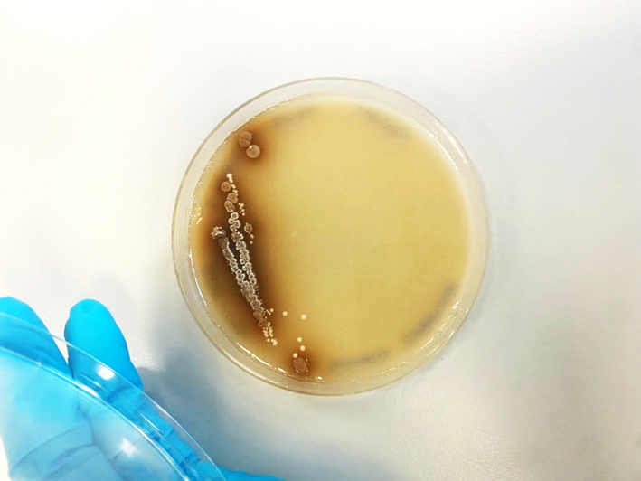 | 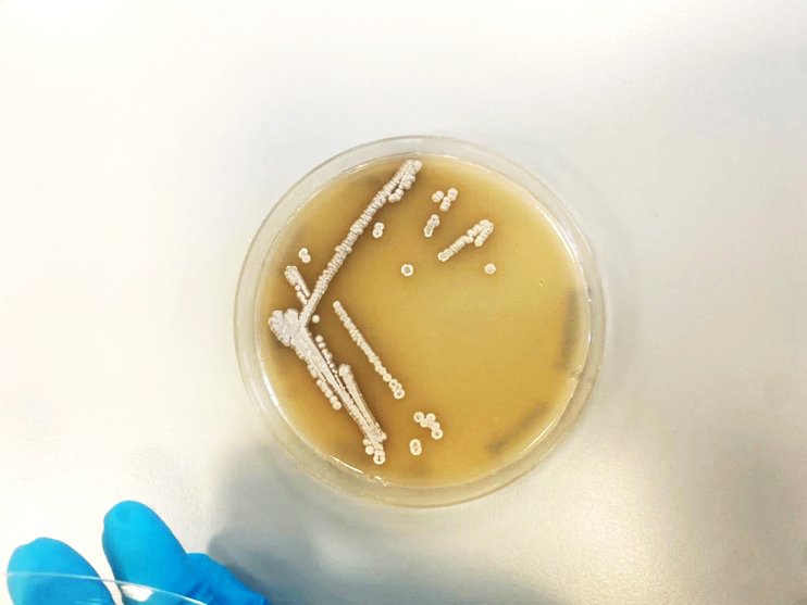 |
| Sample 1 colony streak | Sample 2 colony streak |
Figure 1: Two different suspected Streptomyces soil isolates streaked on SFM plates
- Spreading on spore forming plates
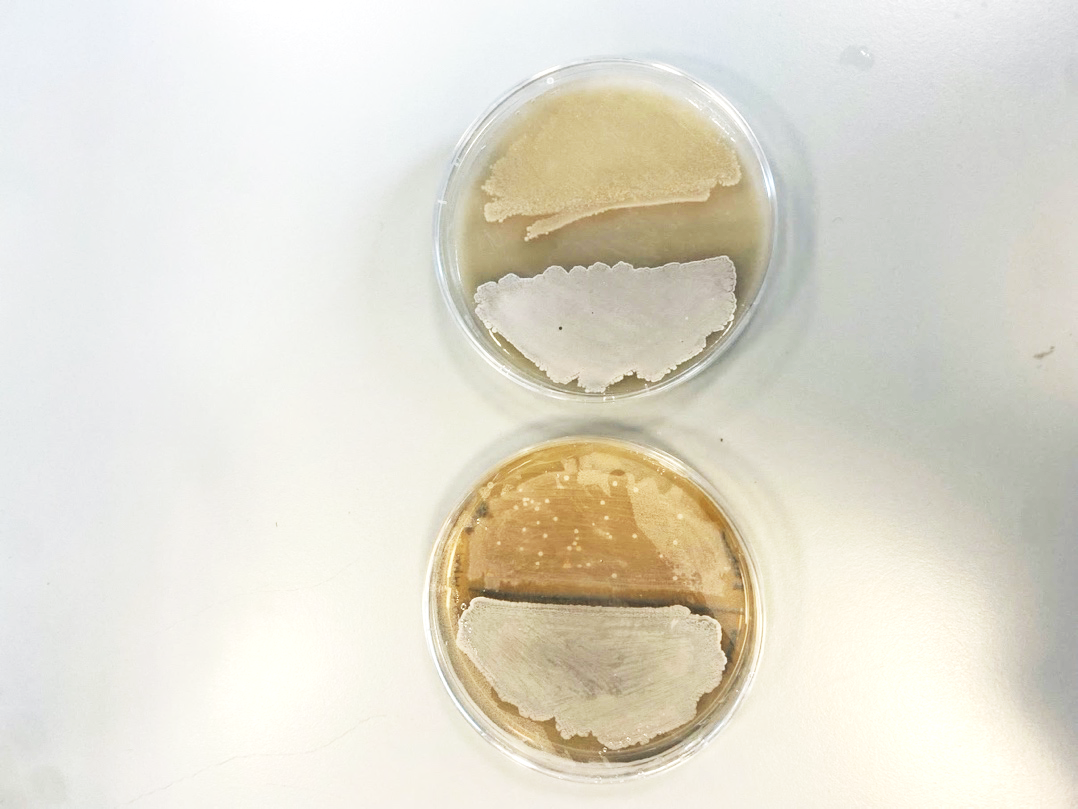 |  |
| SFM plate | MYM plate |
Figure 2: Colony 1 and colony 2 soil isolates plated on SFM and MYM sporulation plates
- Agar plug diffusion test
Table 2: Inhibition zone on indicator bacteria plates by agar plugs from soil samples
| AntibioticBacteria strain | Agar plugs from SFM media | Agar plugs from MYM media | ||
| Soil isolate #1 | Soil isolate #2 | Soil isolate #1 | Soil isolate #2 | |
| M. luteus | 0 | 0 | 0 | 0 |
| E. coli DS941 | 0 | 0 | 0 | 0 |
| E. coli DS941 PSB1C3 | 0 | 0 | 0 | 0 |
Results of plug diffusion test show that none of the soil isolates have produced sufficient antibiotics to inhibit any of the indicator bacteria. There was no inhibition zone in all plates.
- 16S rRNA amplification and visualizing
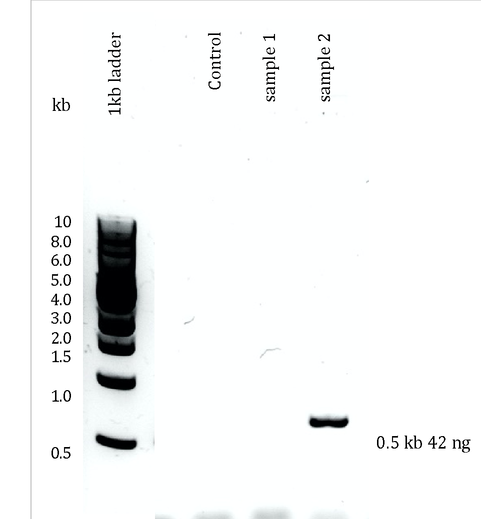
Figure 3: 1% agarose gel for 16S rRNA gene from sample 1 and sample 2, no-templet control, and 1kb ladder.
Gel electrophoresis results show no bands in control and sample 1. There is only one clear intense band for sample 2 of size 0.5 kb and 42 ng indicating that 16S rRNA was successfully amplified by PCR, and thus this sample was chosen for sequencing.
- 16S rRNA sequencing
The concentration of sample 2 16S rRNA sent for sequencing was 16.2 ng/μL, with a good purity value of 1.6.
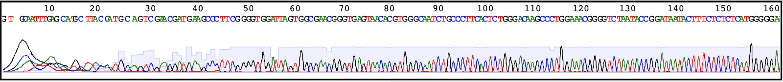
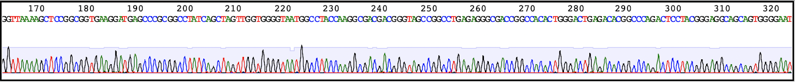
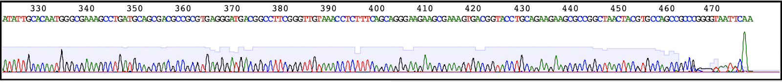
Figure 4: sequence of soil sample 2 as obtained by sanger sequencing, with the location of reverse primer marked at the end of the sequence. And two red lines pointing at the sequence looked at in BLAST database
According to the results obtained from gel electrophoresis (figure 3) the expected fragment size is ~500bp. And The sequence obtained in figure 4 is around the size expected, which is few bases >470. There is 519 Reverse primer sequence seen at the end of the sequence.
Table 3: Sequences producing most significant alignments
| Description | Query Cover | % Identity |
| Streptomyces brevispora strain BK160 16S ribosomal RNA, partial sequence | 100% | 99.50% |
| Streptomyces laculatispora strain BK166 16S ribosomal RNA, partial sequence | 100% | 99.25% |
| Streptomyces drozdowiczii strain NRRL B-24297 16S ribosomal RNA, partial sequence | 100% | 98.75% |
| Streptomyces drozdowiczii strain NBRC 101007 16S ribosomal RNA, partial sequence | 100% | 98.75% |
| Streptomyces sannanensis strain NBRC 14239 16S ribosomal RNA, partial sequence | 100% | 98.25% |
| Streptomyces caldifontis strain NCCP-1331 16S ribosomal RNA, partial sequence | 100% | 98.00% |
| Streptomyces paludis strain GSSD-12 16S ribosomal RNA, partial sequence | 100% | 98.00% |
| Streptomyces olivoviridis strain NBRC 12897 16S ribosomal RNA, partial sequence | 100% | 98.00% |
| Streptomyces atroolivaceus strain NBRC 12741 16S ribosomal RNA, partial sequence | 100% | 98.00% |
| Streptomyces atroolivaceus strain LMG 19306 16S ribosomal RNA, partial sequence | 100% | 98.00% |
As shown in table 3 BLAST nucleotide database cover 100% of the sequence query, it’s indicated that the sample is from Streptomyces species. The most significant alignment is Streptomyces brevispora strain BK160 16S rRNA. Looking more in detail, thre is 99% matching in the alignment of soil sample sequence with its best match Streptomyces brevispora, there are two mismatches, one is a single nucleotide deletion at position 16, and the second is at position 112 C>T.
- Chloramphenicol from wild-type and genetically modified Streptomyces Venezuela


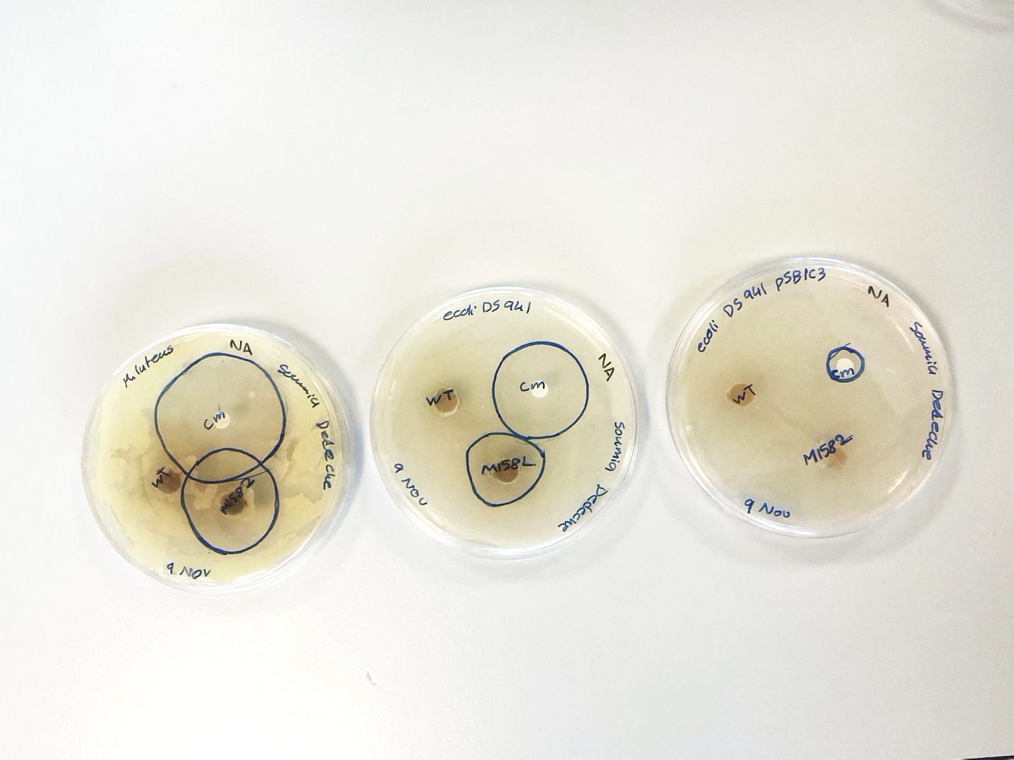
A B C
Figure 5: Three indicator bacteria plates: (A) M. luteus (B) E. coli DS 941 (C) E. coli DS 941 PSB1C3. Showing zone of inhibition as an action of Cm control antibiotic disc, wild-type S. venezuelae, agar plug and M158L S. venezuelae agar plug.
Table 4: Inhibition zone by agar plugs assays against indicator bacteria
| AntibioticBacteria strain | Cm | M1582 | wild-type |
| M. luteus | 42 mm | 35 mm | 0 mm |
| E. coli DS941 | 32 mm | 24 mm | 0 mm |
| E. coli DS941 PSB1C3 | 11 mm | 0 mm | 0 mm |
Cm control discs caused the highest inhibition compared to the agar plugs of S. venezuelae. S. venezuelae M1582 plug strain inhibited M. luteus and E. coli DS941 but not E. coli DS941 PSB1C3. The wild-type S. venezuelae did not produce sufficient antibiotics to inhibit any of the indicator bacteria.
- Discussion
- Antibiotics from soil samples
Looking at table 1, It is shown that the undiluted sample in plate Cyc+Nal 100 have the highest number of bacteria, however, very few of them are suspected Streptomyces. Cyc plates have higher number of colonies for the same dilutions compared to the Cyc+Nal plates, which is due to the selectivity of nalidixic acid that eliminates most gram-negative bacteria (Crumplin & Smith, 1975), Cycloheximide plate is less selective, since Cycloheximide only inhibits fungal growth (Bradley, 1962). Overall, the number of suspected Streptomyces was only 3 colonies from Cyc+Nal non-diluted sample, and 3 from 10-1 Cyc sample. Two of the suspected colonies streaks obtained pure colonies (figure 1). Morphological characteristics of the two colonies were almost the same, both are white hard, powdery dry looking, however, they have few morphological differences as colony 1 is darker and have brown secretion.
SFM and MYM sporulation plates (Figure 2) show an even distribution of samples from colony 1 and colony 2, It is also shown that colony 1 in MYM produced some white dots which might be due highly intense bacteria spots. Agar plug diffusion test for both samples presented in table 2 shows no inhibition in all the indicator bacteria. Which indicate that the species isolated from soil did not produce efficient antibiotics to kill the indicator bacteria. Although most streptomyces species produce variety of secondary metabolites and antibiotics in nature, however, more optimized standardization of soil sample selection, and more trials might be required to find significant antibiotic production.
Amplification of 16S rRNA gene from soil isolates was visualized in gel electrophoresis (Figure 3). The non-templet control shows no band indicating no contamination. Sample 1 shows no band which indicates that primers used for amplification didn’t work on this sample and therefore it might not be the targeted Streptomyces. However, sample 2 showed a band of size 0.5 kb and 42ng. This band indicates that 16S rRNA has been successfully amplified from this sample. Therefore, it was sent for sequencing identification.
The comparison of 16S rRNA sequence in BLAST database indicates that it is related to Streptomyces species, as all alignment sequences present in the database are for Streptomyces. Moreover, the results suggest that the sample is related to Streptomyces brevispora due to its high similarity with Streptomyces brevispora strain BK160 16S r RNA. With very few mismatches between the query and Streptomyces brevispora strain sequence. These results indicate that the experiment has successfully isolated Streptomyces from the soil, however, it didn’t produce significant antibiotics to inhibit indicator bacteria.
- Chloramphenicol from Streptomyces Venezuela
Inhibition zones marked by circles in figure 5 indicate the effectiveness of chloramphenicol in some samples. The circles diameter was measured and recorded in Table 4. Which shows that control Cm have the highest chloramphenicol efficiency, followed by S. venezuelae M1582, and the least effective among plugs was the wild-type S. venezuelae which didn’t inhibit any of the indicator bacteria. On the other hand, among indicator bacteria, E. coli DS941 PSB1C3 was the most resistant as it carries PSB1C3 chloramphenicol resistance gene (Cmr). Then E. coli DS941 strain shows moderate resistance, and M. luteus shows the least resistance level. These results confirm that the effect of enhanced expression of sven0913 gene in the genetically modified S. venezuelae M1582 upregulates chloramphenicol gene cluster, hence, successfully enhances chloramphenicol productivity.
- References
de Lima Procópio, R. E., da Silva, I. R., Martins, M. K., de Azevedo, J. L., & de Araújo, J. M. (2012). Antibiotics produced by Streptomyces. The Brazilian Journal of Infectious Diseases, 16(5), 466–471. https://doi.org/10.1016/j.bjid.2012.08.014
Bradley, S. G. (1962). Relationship between Sugar Utilization and the Action of Cycloheximide on Diverse Fungi. Nature, 194(4825), 315–316. https://doi.org/10.1038/194315a0
Colloms, S. (2021). A Laboratory Project Investigating Production of Antibiotics by Streptomyces. Industrial and Environmental Microbiology Lab – MSc Biotech 2021/22.
Crumplin, G. C., & Smith, J. T. (1975). Nalidixic Acid: an Antibacterial Paradox. Antimicrobial Agents and Chemotherapy, 8(3), 251–261. https://www.ncbi.nlm.nih.gov/pmc/articles/PMC429302/
D’Costa, V. M., Griffiths, E., & Wright, G. D. (2007). Expanding the soil antibiotic resistome: exploring environmental diversity. Current Opinion in Microbiology, 10(5), 481–489. https://doi.org/10.1016/j.mib.2007.08.009
Fernández-Martínez, L. T., Borsetto, C., Gomez-Escribano, J. P., Bibb, M. J., Al-Bassam, M. M., Chandra, G., & Bibb, M. J. (2014). New Insights into Chloramphenicol Biosynthesis in Streptomyces venezuelae ATCC 10712. Antimicrobial Agents and Chemotherapy, 58(12), 7441–7450. https://doi.org/10.1128/aac.04272-14
Huemer, M., Mairpady Shambat, S., Brugger, S. D., & Zinkernagel, A. S. (2020). Antibiotic resistance and persistence—Implications for human health and treatment perspectives. EMBO Reports, 21(12). https://doi.org/10.15252/embr.202051034
Hutchings, M. I., Truman, A. W., & Wilkinson, B. (2019). Antibiotics: past, present and future. Current Opinion in Microbiology, 51(1), 72–80. https://doi.org/10.1016/j.mib.2019.10.008
Nicolaou, K. C., & Rigol, S. (2017). A brief history of antibiotics and select advances in their synthesis. The Journal of Antibiotics, 71(2), 153–184. https://doi.org/10.1038/ja.2017.62
Zaffiri, L., Gardner, J., & Toledo-Pereyra, L. H. (2012). History of Antibiotics. From Salvarsan to Cephalosporins. Journal of Investigative Surgery, 25(2), 67–77. https://doi.org/10.3109/08941939.2012.664099